Medical imaging, and the evidence that says when the model is wrong.
Applied ML research engineer, PhD. Two recent pieces of work, both on public data, both with the negative results left in.
Endoscopy · acquisition shortcuts
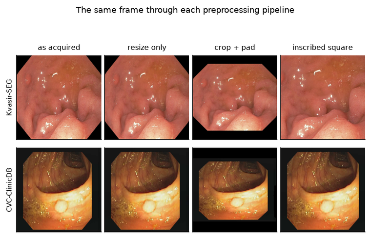
You can tell which hospital a colonoscopy frame came from 96% of the time, without looking at the anatomy — and the standard fix barely helps.
Nine numbers describing the frame the equipment drew — letterboxing, the field-of-view mask, the burned-in date — identify the source dataset out of five, at chance 0.20.
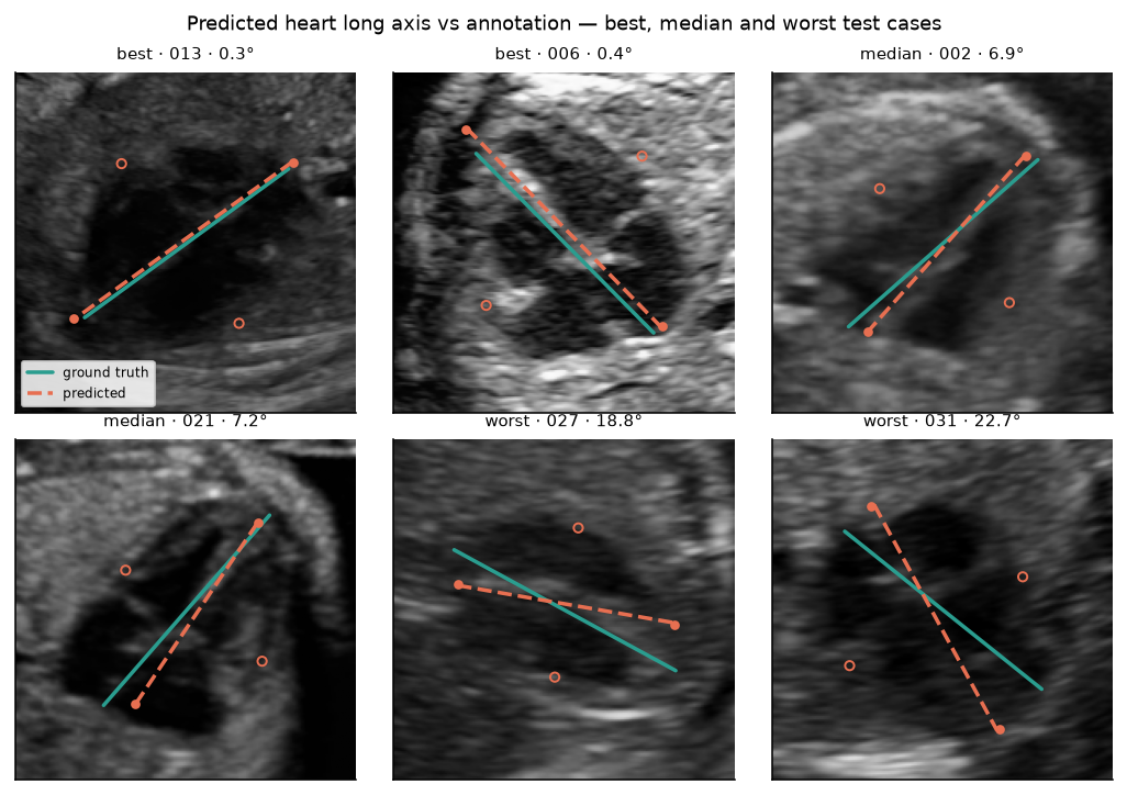
Estimating fetal cardiac orientation does not need a trained model — closed-form geometry beats the network by two orders of magnitude.
The useful part is knowing when that shortcut breaks. It does not break where the usual quality score says it should: a mask scoring Dice 0.87 can give a 46° error, one at 0.77 gives 0.22°.
Previously: self-supervised pretraining of a medical-imaging foundation model on a large clinical video corpus under NDA, multi-GPU — with the cross-vendor standardisation and evaluation harness underneath it. Before that, seven years of computer vision and robotics in industry in Germany.